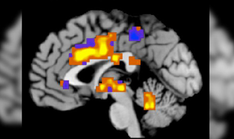
This image shows regions of the "neurologic pain signature," a standard map that can be applied to individual people who may be experiencing pain (Photo: AP)
Scientists have finely sliced a human brain into 7,400 wafer-thin sheets and then reconstructed it to create the world's most detailed map of the brain in three dimensions.
The so-called "Big Brain" project, which took a 65-year-old woman's brain and cut it into slices just 20 micrometres thick, shows the brain's anatomy in microscopic detail, almost down to a cellular level, said the scientists who carried out the work. A micrometre, or micron, is a millionth of a metre.
The resulting 3D reference brain will allow researchers worldwide to study the organ in unprecedented physical detail, paving the way for deeper understanding of how processes like cognition and emotions work, and how brain diseases develop.
"This is big science comes to the brain," said Dr Alan Evans, a professor at the Montreal Neurological Institute at McGill University in Montreal, Canada, who co-led the study.
"We've raised the level of insight (by) orders of magnitude," he added. "This data will revolutionise our ability to understand internal brain organisation."
Although neuroscientists have created reference brains in the past and used them to study how physical form relates to the brain's chemical signalling and genetic makeup, this is the first 3D brain map to offer such detail - at a spatial resolution of 20 microns, smaller than the size of one fine strand of hair.
Until now, scientists had only been able to study the brain on a scale of about 1 millimetre by 1 millimetre by 1 millimetre, Evans told reporters in a teleconference.
"We're now 50 times smaller in all three dimensions," he said. "This changes the game in terms of our ability to discriminate very fine structural and physiological properties of the human brain."
Because the sliced and reconstructed Big Brain was created from a dead organ, scientists said the best way to imagine it was as a type of scaffold providing a 3D framework for living brain information to be analysed.
Using a special tool called a microtome, the researchers from Canada and Germany sliced the 65-year-old human female brain, which had been embedded in paraffin wax, into more than 7,400 sections.
They then mounted the 20-micrometre thick sections onto slides, stained them to detect cell structures and then digitised them with a high-resolution scanner so that the 3D brain model could be reconstructed. It took around 1,000 hours to collect the data.
The fine-grained anatomical resolution should give scientists insights into the neurobiological basis of cognition, language, emotions and other brain processes.
"The (researchers) pushed the limits of current technology," said Peter Stern, editor of the journal Science where the work was peer-reviewed and published on Thursday.
Short link: